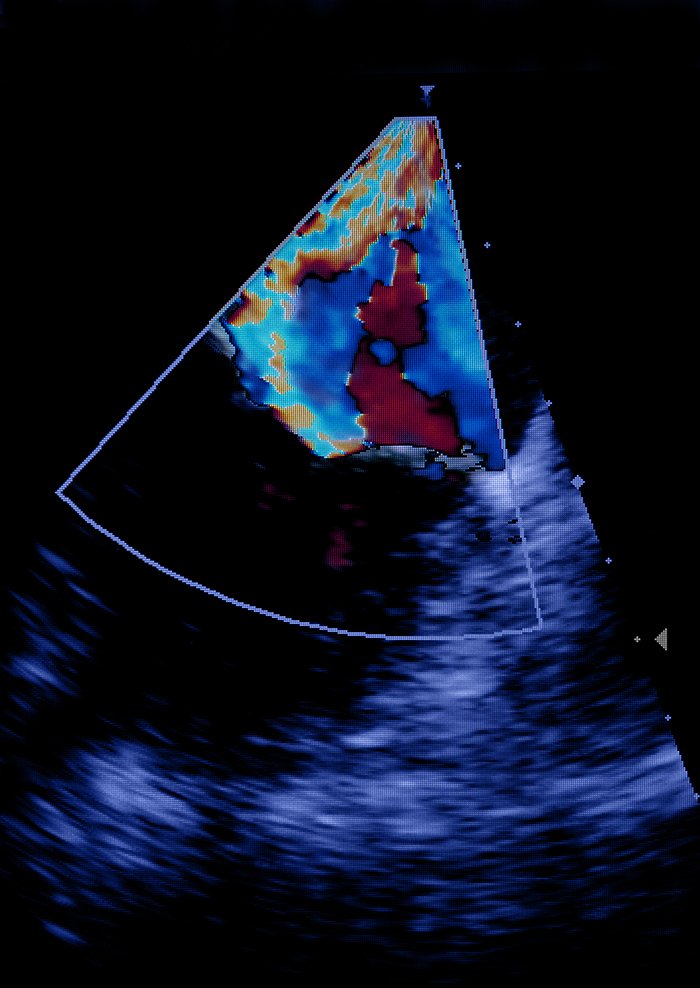
Contrast Echocardiography
It is estimated that up to 20% of resting transthoracic echocardiograms have suboptimal left ventricular endocardial visualization, defined as two or more segments not being adequately visualized. This limitation underscores the importance of high-quality imaging for accurate diagnostic results.
Overview
Contrast Echocardiography, using Definity as a contrast agent, enhances the sensitivity of standard transthoracic echocardiography (TTE) by improving the visualization of cardiac structures and blood flow, particularly in patients with suboptimal baseline images. This procedure is especially beneficial when traditional echocardiography fails to provide clear delineation of myocardial borders, ventricular wall motion, or the endocardial surface, enabling more accurate diagnostics.
Clinical Indications for Contrast Echocardiography
Chest pain evaluation
Hypertrophic cardiomyopathy
LV ejection fraction and regional wall motion assessment
LV thrombus and intracardiac mass evaluation
LV aneurysm versus pseudoaneurysm
LV noncompaction
Other less common apical abnormalities
Intracardiac abnormalities
Suboptimal left ventricular endocardial visualization
Additional Clinical Indications for Contrast Echocardiography
Left Ventricular Dysfunction: In patients with poor acoustic windows or inadequate image quality, contrast administration significantly improves left ventricular endocardial border delineation, aiding in more accurate assessments of function and contractility.
Assessment of Valvular Disease: For improved visualization of valvular regurgitation, particularly in cases with suboptimal Doppler signals, contrast echocardiography enhances the detection and quantification of regurgitant jets.
Suspected Adult Congenital Heart Disease (ACHD): When assessing complex congenital defects, contrast helps identify abnormal communications, shunts, and residual defects.
Evaluation of Endocarditis: In cases of suspected infective endocarditis, contrast assists in visualizing vegetations, especially when standard echocardiography provides limited visualization.
Aortic Root and Marfan's Syndrome: Contrast offers better visualization of the aortic root, which is critical for evaluating conditions like Marfan's syndrome.
Procedure
Definity, a perflutren lipid microsphere contrast agent, is administered intravenously to enhance echocardiographic images. The microbubbles reflect ultrasound waves, increasing contrast between the blood pool and surrounding tissues. This agent improves the clarity of left ventricular endocardial borders, valvular anatomy, and blood flow dynamics, facilitating more accurate assessments.
Possible Side Effects
Definity is generally well-tolerated. However, potential side effects include:
Mild Reactions: Headache, dizziness, nausea, flushing, back pain, or mild chest discomfort.
Injection Site Reactions: Minor bruising or infection at the IV site.
Serious Reactions: Rare but potentially serious reactions, such as hypersensitivity, respiratory distress, or arrhythmias, occur in fewer than 0.01% of patients (approximately 1 in 10,000 cases), typically within 30 minutes of administration. Close monitoring post-administration is essential in these cases.
Contraindications
Hypersensitivity to perflutren or albumin
Acute coronary syndrome or unstable heart failure
Patient Considerations
Please inform the sonographer if the patient has any of the following conditions:
Acute myocardial infarction or acute coronary syndrome
Unstable or worsening congestive heart failure
Serious ventricular arrhythmias
Respiratory failure
Pregnancy or breastfeeding
Procedure Monitoring
Patients with pre-existing conditions will be monitored for approximately 30 minutes following the administration of Definity.
Report Processing
The contrast echocardiography report is meticulously analyzed by a cardiologist. A detailed report, including conclusions and clinical recommendations, is sent directly to the referring physician. This ensures timely and accurate follow-up care.
Next Steps
To refer a patient for Contrast Echocardiography or any other diagnostic modality, please download the Requisition Form and fax it to our Requisition Processing Centre at 647-351-6648.
References
Baumgartner, H., Falk, V., Bax, J. J., et al. (2017). Cardiovascular management of adults with Marfan syndrome. European Heart Journal, 38(20), 1543-1579. https://doi.org/10.1093/eurheartj/ehw614
Fernández-Golfín, C., Fernández-Golfín, J., López Fernández, T., et al. (2011). Evaluation of congenital heart disease in adults using contrast echocardiography. Revista Española de Cardiología, 64(9), 789-796. https://doi.org/10.1016/j.recesp.2011.06.006
Porter, T. R., Xie, F., Kricsfeld, D., & Kilzer, K. (1997). Detection of cardiac sources of embolism using contrast echocardiography. Circulation, 95(6), 1686-1691. https://doi.org/10.1161/01.cir.95.6.1686
Senior, R., Monaghan, M., Becher, H., & Nihoyannopoulos, P. (2005). Contrast echocardiography: Evidence-based recommendations by European Association of Echocardiography. European Journal of Echocardiography, 6(1), 3-15. https://doi.org/10.1016/j.euje.2004.09.001